Tag Archives: environmental microbiology

In the spirit of the day…

14 Feb

Despite the fact that I really don’t celebrate VD (Valentine’s Day, in case you were thinking something else), I thought I would show a little love, share a warm-fuzzy, and re-post this from Suzanne Kennedy (clearly, a woman after my own heart) at the MoBio Blog,  The Culture Dish.

Oh how do I love thee? Let me count the ways…

Show some LOVE for Environmental Microbiology

Do you love your work? Does nothing make you happier than a day out in the field collecting soil from the rainforest floor, in a boat collecting Vibrio contaminated water from Puget Sound, traipsing the forest looking for animal droppings from wild birds in Venezuela, or aboard the Alvin collecting biofilms from deep sea floor hydrothermal vents?

It’s important to love your work and fortunately for us, there is so much to love about microbiology and the environment. But to find out what is best about working in this field, I asked the question to several of my scientist friends:  What do you love about your work? Why do you study environmental microbiology and what is it that makes it the best field of work?

And below are some of the best responses. Some are my own, but most are responses I received from people who study some of the most unusual samples from the most extreme environments in the world.  I think you will agree that environmental microbiology provides experiences unlike any other field. Let us know your reasons for loving your work!

14 Reasons to Love Environmental Microbiology:

1. You get to play outside in the mud, snow, water or clouds (see picture at end of article).

2. There is virtually an unlimited number of research projects to choose from. “Microbiologist William B. Whitman, estimates the number of bacteria in the world to be five million trillion trillion. That’s a five with 30 zeroes after it. Look at it this way. If each bacterium were a penny, the stack would reach a trillion light years.”

3. Your research will have an impact on everything living on the planet, humans, animals, and plants. Basically all the Kingdoms benefit from what you do.

4. You have the opportunity to visit exotic and remote locations.  

Graduate student Rick Davis explains, 

“I think I’ve been really lucky with the places I get to study– I got to go to Samoa, Hawaii, and Yellowstone this year!”

He also added reason number 5: 

5.  Environmental microbiologists are more laid back and generally more collaborative than competitive, which allows for greater progress and more fun at conferences!

John Mackay, a molecular biologist and director of business development at the plant diagnostic company, Linnaeus, tells me:

6. You can cruise around the seas for months, sequence a bit of sea water and write the whole lot off on your research grant!

7. You can work on things you can eat or drink – I recommend wine and truffles!

8. When you find new species (almost a given!), you can name them after yourself.

 New discoveries are also what motivates Charlie Lee from the University of Waikato, a Postdoctoral researcher in microbial ecology studying the Dry Valleys of Antarctica. He echoes the sentiment that discovery is almost a guarantee:

9. Most systems we look at are relatively poorly understood, and it’s always exciting to discover something for the first time.

 Tom Niederberger, a Postdoctoral researcher in marine biosciences at the University of Delaware, has more to add:

10. The international travel is a great reward. The world is your playground as microbes have colonized basically all habitats on earth, and it’s great to travel around sampling not only the microbes, but new cultures/food/travel etc. and not being chained to the lab and pipette. Also the international collaborations and conferences also are great.

11. But I think what is most important is that microbes in the environment are essential not only for the health of the planet (e.g. global nutrient cycling / global climate change) but they are also intimately linked to the healthy functioning of our bodies. i.e. the are really important!

12. Also,there is the excitement of the unknown. Most of the organisms cannot be cultured and we know nothing about them…I think this is great motivation and it will keep you busy, and there are always new problems to solve and new questions to ask.

All excellent points!

And from a molecular biologist from Colorado State University (who wished to remain anonymous) come two excellent points I hadn’t considered:

13. Extremists don’t kidnap environmental microbiologists. Actually, they give them back.
 
14.
If you get tenure, who’s going to boot you out?  Exxon?

Did I mention that environmental microbiologists are funny?

Helper Microbes and Heavy Metals…

3 Sep
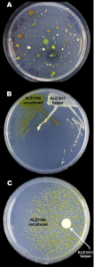
Marine bacteria exhibiting "helper" facilitated growth.

Series of Petri plates with marine bacteria exhibiting "helper" facilitated growth of previously uncultured organisms.

Last week, the American Society for Microbiology posted a story that caught my eye which highlighted the most recent work of Kim Lewis and his collaborators published in Chemistry and Biology in March (see citation below).  It caught my eye due to the term “siderophores” in the title.  You may be wondering why a strange Greek term like that would be of any interest to a soil microbiologist like me, so let me share my fascination for these weird and wonderful molecules and how they may be changing approaches in microbiology.

First, let’s define the word siderophore in simple language. 

It’s from the original Greek meaning “iron carrier” and describes a small molecule specifically designed to address the biological iron requirement.  While you and I might be able to eat our Flintstones and therefore don’t need to worry about iron, the vast majority of life on earth has to figure out a way around the fact that biological systems require the soluble Fe2+ (ferrous iron), but iron for the most part exists in the environment as the highly insoluble Fe3+ (ferric iron).  Why?  Because life evolved when the earth was anaerobic (meaning there was virtually no oxygen); there was an overabundance of iron all over the place in the ferrous form.  Gradually life took over the face of the planet, turning out atmosphere aerobic, and all that oxygen essentially ‘spoiled’ the iron-rich crust of the earth, oxidizing the iron into the ferric form, which is essentially insoluble and not bioavailable. 

Long-story short, we’ve all developed some strategy to deal with this problem.  Humans use transferrin in our bloodstream to bind iron, while bacteria, fungi, and some plants use what we call siderophores.  These small molecules grab onto ferric iron in the environment and chelate it, forming soluble iron complexes, making it available for biological purposes.  For a really gre,at in-depth review of siderophores, look here.

Curiouser and curiouser…

What has intrigued me for years about siderophores is a uniquely discriminating selectivity.  I know that may sound redundant, but what I’m trying to say is that certain microbial siderophores are extremely specific to iron and actually can scavenge iron from metamorphic rocks, oxides, hydroxides, you name it they can leach it out, to the point that they even have biotechnological applications in mining activities.  At the same time, other microbial siderophores may be able to pick up any positively charged metal or micronutrient in the environment.  This includes all the common heavy-metal contaminants that I deal with on a daily basis, from lead and cadmium, to arsenic and uranium.  Certain siderophores can chelate these toxic metals and bind them in such a way as to allow them to precipitate out of solution, making them no longer a toxic threat to the organism.  Even more fascinating is that the same siderophore, under different conditions, might bind a scarce essential nutrient so the microbe can take it up and continue living normally.  There has also been evidence recently to support that microbial siderophores may be involved in quorum sensing (microbial cross-talk) and biofilm formation, but the new work by Kim Lewis and his colleagues brings it all to a new level.

The “great plate county anomaly”

For years, microbiologists have been working on new ways to culture (or, grow in the lab) environmental microorganisms and attempts to overcome the “great plate count anomaly” have included a multitude of creative approaches.  All my microbiologist-readers will be intimately familiar with that little problem, but for those of you out there who aren’t … the “great plate count anomaly” describes the fact that no matter how many microbes we can see under a microscrope or detect DNA for in any given sample (soil, marine sediment, water, mucosa, etc), we can only successfully get around 0.1 – 1% to grow on Petri plates in the lab.  This severely limits what we can find out about how these organisms function out in the real world and has stalled many aspects of microbial ecology.

The authors were able to definitively prove the siderophores produced by some “helper” microbes actually allowed the growth of other, previously uncultured, bacteria on Petri plates in the lab.  They were able to identify cooperative pairs of organisms, in which one organism was identified as the helper and secreted siderophores into the media, and the other organism was only capable of growth when exposed to the siderophores of the helper.  They tried an array of synthetic (store-bought) siderophores, which worked in some cases but others did not. They also tried to supplement with high quantities of bioavailable iron, which also, worked in some cases (allowed the growth of previously uncultured microorganisms), and in others did not. This means the helpers likely have very specific relationships with the organisms that rely on their siderophores, with a great degree of discrimination, despite the fact the pairs of organisms were not closely related, not even in the same genus.  It may also mean that the function of the siderophore in the media may not have anything to do with iron in some cases.  By using these helper organisms the authors were actually able to culture as much as 40% of the total community, which is a vast improvement of the typical <1% we see from most environmental samples.

As a scientist who has literally watched this phenomena unfold on my own Petri plates, but had no clear explanation at the time, I am truly enthralled by this new discovery.  It raises some very interesting questions with regard to the microbial ecology of this helper-pair system, in addition to opening doors with regard to antimicrobial therapies and basic culture techniques.     

But, I have to wonder, why?  What’s the purpose of this highly complex and specific signaling and nutrient acquisition cross-talk?  How would it benefit the microbial community as a whole?  Is this cooperation and microbial co-evolution at its best, right before our eyes?  Things I’ll be pondering over the weekend…

________________________________________________________________________________________________________________________________________________________________________
ResearchBlogging.orgD’Onofrio A, Crawford JM, Stewart EJ, Witt K, Gavrish E, Epstein S, Clardy J, & Lewis K (2010). Siderophores from neighboring organisms promote the growth of uncultured bacteria. Chemistry & biology, 17 (3), 254-64 PMID: 20338517

Bioremediation with Beeswax

13 Jul

Two years ago, I showed this video in a class I was teaching on Environmental Microbiology and I still love it… it’s a fantastic example of biostimulation that I just had to share when I saw a story on  the possible application of PRP in clean-up efforts in the Gulf

A NASA partnership in the early 1990’s created a fine powder out of beeswax, called PRP, which can be spread over petroleum or oil to absorb (or encapsulate) the oil at the same time providing essential nutrients (nitrogen, phosphorus, and potassium) to the oil-eating microbes and speeding up the break-down process. 

Gotta love the History Channel!

Toxic Tradeoff: An Oil-Dispersant Story

12 Jul

I’ve been interested in the discussions on oil dispersants being used in the BP gulf disaster recently, specifically from the microbial break-down perspective.  Theoretically, dispersing the oil should give it greater surface area, allowing more rapid chemical and biological degradation rates. Speedy biodegradation seems like a good thing.   However, I just discovered an article published this spring in the International Journal of Environmental Research   which gave me pause to think on the whole issue in a way I hadn’t before. 

The article’s author (Dr. A. Otitoloju – a research scientist in the department of Zoology at the University of Lagos, Nigeria ) evaluated crude oil break-down under natural, versus dispersant-controlled settings and the findings seem especially meaningful in light on the current situation in the gulf. First, I should mention that this subject area is much more toxicology than microbiology and therefore my knowledge base is limited.  So it was particularly interesting for me to find out from this article that, “most of the dispersants that have been initially introduced into the market were found to either to be very toxic on their own or enhance the toxicity of the spilled oil on the receiving habitat or exposed organism when deployed to control oil spills.”  Of course this isn’t news to everybody and BP’s use of the toxic COREXIT ® has been the subject of some heated debate already  mostly due to the fact that there are  other dispersants tested by the EPA  found to be less toxic and more effective.

Perhaps it isn’t surprising then that this research article by Otitoloju found increased toxicity of the oil-dispersant mixture to the test organisms, African catfish.  But what is interesting is that over the 28 days of the study, the level of toxicity did not decrease but actually increased over time.  Overall, the dispersant mixture was about twice as deadly to the fish as was the uncontrolled oil, mostly likely due to increased exposure (if the oil is in dense clods or balls, or floating on the surface of the water, the fish can’t get as easily coated with and absorb the oil  as it can when the oil has been chemically dispersed thoughout the water column).

However, it was clear that microbial growth was enhanced (more than 7-fold) and breakdown of the oil was more rapid when the dispersant was used.  So while the dispersant-oil mixture was clearly toxic to the fish (and likely other vertebrates), it enhanced degradation of the oil by microbial populations.  Worth noting, though, are the facts that:

1.) Even without dispersant added, the oil did begin to break down over the course of the 28-day study and gradually decreased in overall toxicity to the fish; and,

2.) Inreased microbial breakdown may be short-lived, according to an article in the ScienceInsider which discusses dispersant interference with natural microbial processes in marine systems.

Now, in thinking about how this pertains to the Gulf of Mexico, where almost 2 million gallons of dispersant have been applied already (and more every day), how will this play out in the long-run?  The dispersant makes the oil less obvious floating on the surface of the water and reduces the appearances of “tar-balls” on beaches, but increases bioavailability and exposure of the wildlife significantly, resulting in higher levels of toxicity, despite [temporarily]  increased microbial degradation rates.

So what exactly is the trade-off? 

Dispersant use equates more dead fish, coral, and so on, in the short-term, but with more rapid disappearance of the oil from the waters and coastline in the long-run (see also the article by M. Torrice cited below).

Degradation rates might be even more critical when we consider hurricane season could bring substantial quantities of oil inland. Maybe a dispersant is a necessary evil.  But in that case it seems only right to use the least toxic, most effective dispersant available, which certainly isn’t COREXIT ®.

(For more toxicity info, see the EPA’s Toxicity Testing of Dispersants for the Gulf, last updated June 30th)

________________________________________________________________________________________________________________________________________________________________________

Otitoloju, A.A. (2010) Evaluation of crude oil degradation under a no-control and dispersant-control settings, based on biological and physical techniques International Journal of Enivronmental Research, 4(2): 353-360.

Torrice, M. (2010) Cleaning up the Gulf oil spill.  Chemical & Engineering News, 88(20): 36-37.

The Newest Bacterium: Part 3, Finalé

6 Jul

How did the Venter research group synthesize a genome for transplant? 

The first thing I really wanted to know in reading the original research article in Science (May 20th, 2010) was, how did the authors of this ground-breaking piece of science manage to synthesize an entire bacterial genome? 

And the answer is: they didn’t, not exactly. 

If you’d like to see for yourself, the actual materials and methods used in the Science article were not contained within the article itself.  They are, however, available here as “Supplementary Materials.” 

But if you’d rather not sit down with your molecular microbiology text book and dictionary to go through their 29 pages of methods, let me explain as briefly as possible…

As I mentioned in the previous post on this (Part 2), Venter’s group of scientists at the J. Craig Venter Institute (JCVI) had already worked with these Mycoplasmas for quite some time and had sequenced at least one entire genome.  In doing so, as well as a series of experiments in which they could delete one gene at a time and see what happened to the bacterium, they managed to figure out precisely which genes were necessary for survival in the lab and which were not.

At that point they simply ordered the genome sequence, in little pieces, from a company called Blue Heron  that commercially manufactures DNA in the form of oligonucleotides (a.k.a. oligos); an oligo is a small piece of DNA arranged according to your (consumer) specifications. 

There are a variety of companies in existence all over the world that will put together oligonucleotides for any researcher or institution; you only have to tell the company what order you want the bases in (A, T, C, and G).  Venter and the scientists at the JCVI simply ordered the known sequence of the genome of M. mycoides from Blue Heron in pieces each about 1,080 bases long, and made sure that the pieces all had overlapping, matching sequence at the ends. 

So, the Venter group also wanted to make sure that their soon-to-be creation would not only function properly, but could be identified, patented, and its movement tracked.  Therefore, in writing this synthetic genome, they included some “watermark” non-coding sequences.  These are bits of genetic material that don’t actually get used in the daily life-functions of the bacterium; they are just there, taking up space, allowing recognition and quality assurance by the scientists working with the organism.  In these watermark sequences the scientists spelled out their own names, a few choice quotes, and even a webpage, all in the genetic code of this organism (more info on the code at another interesting blog).

But how did they assemble the pieces of DNA into a genome?

Two words:   slave   labor 

Only, not the way you might be thinking.  They used a common yeast (Saccharomyces cerevisiae; a.k.a. “bakers’ yeast” or “brewers’ yeast”) to put all the pieces together for them.  This particular yeast was the first fully sequenced eukaryotic (eukaryote = cells with a true nucleus, i.e. plants, animals, us, not bacteria) organism and is particularly easy to grow. 

Since its genome was sequenced more than ten years ago and it’s the “model organism” for eukaryotic cell function, we know a lot about it, how it works, but more specifically, how it’s DNA repair and synthesis mechanisms function. 

Dr. Venter and his team of scientists made use of this knowledge in the way they designed the pieces of the bacterial genome that they were interested in transplanting.  There were specific recognition sites allowing the natural machinery of the yeast cell to stitch them all together in a specific order.  They had to go through three rounds of yeast-facilitated patch-work-quilting to get the whole genome, starting with the small pieces they ordered from Blue Heron, then gradually making larger and larger pieces until one of their yeasts finally contained the entire “synthetic” Mycoplasma mycoides genome (plus watermark sequences).

My 2¢ on the whole thing…

The researchers at JCVI actually published another paper in Nucleic Acids Research only about 3 months earlier describing this entire process, entitled “Cloning whole bacterial genomes in yeast.”  They do a nice job of explaining that microbial gene cloning  has been done for years in microbiology laboratories all over the world for a variety of reasons.  Now, just read that last sentence again slowly and let it really sink in. Then think about this: In 1973, Stanley Cohen and Herbert Boyer actually created the first recombinant DNA (cloned) organism and we’ve been using this technology ever since. (Cloning technology development timeline)

This paper in Science which has caused all this hubbub simply pushed a common method a bit further, from just one gene or a few genes, into the ability to manipulate a very small bacterial genome.  One major driver to this type of research (amongst others) has been the difficulty in not being able to grow most microbes in the lab for detailed study. (The uncultured microbial majority)

If you can’t grow it in the lab, you know fairly little about the organism or what it does out in nature.  We can sequence the same microbe, but still we aren’t certain how that genetic code amounts to functionality in the environment.  The primary advantage to the JCVI’s newest technique over the cloning that we’ve been doing previously is that we can use much larger pieces of DNA, longer gene sequences, and this means more information

The limitations of this technique are obvious when we consider the fact that the transplanted genome was in fact that of Mycoplasma mycoides (with a few additions and deletions).  Not only was the genome specific to that organism, but all the constraints to making this experiment work will be equally as specific.  Try a different genus and the whole ballgame has changed. 

That said, I do acknowledge the fact that these two organisms and the M. mycoides genome may serve as a useful platform or starting place.  I would imagine next steps will be to put together sets of genes from a host of different organisms to create hybrids with the M. mycoides sequence, or some other very simplified genome. In this case, the donors of the genes tacked on to the base genome wouldn’t have to be limited to microbial species,  many genes and gene fragments have already been patented  amid some more recent controversy (roughly 20% of the human genome, in fact).  Each new hybrid, of course, would be patentable in its own right and you can easily see how lucrative this line of research will soon be.

Did I say I’d be brief?

So, in short, the research group at JCVI combined several techniques commonly used in the various fields of microbiology and transplanted the piecemeal genome of one organism into a closely related organism where it began to function rather normally as the first organism.  An interesting and noteworthy piece of science, it is not, in my opinion, the “creation of life” that merits the fear and awe with which the world reacted (at least not yet).

___________________________________________________________________________________________________________________________________________________________________________

 Gibson, D.G. et al. (2010) Creation of a bacterial cell controlled by a chemically synthesized genome.  Science, 329 (5987): 52-56.  DOI: 10.1126/science.1190719

The newest bacterium: Part 2

2 Jul

Myco what?

In the introduction to the article “synthetic genome” article in Science, the authors explain that they had begun work towards this end with Mycoplasma genitalium over 15 years ago. As the name suggests, this bacterium is a human parasitic organism, often found on linings of the urogenital tracts. However, the final, published, “synthetic genome” work used two different Mycoplasma species, M. mycoides and M. carpricolum, neither of which is a human pathogen, but both of which can cause a variety of problems in cattle and goats.

As an environmental microbiologist, I’m not particularly familiar with this genus and I had to ask myself: what’s a Mycoplasma and why use these organisms?

It didn’t take digging too deep to find out that members of the genus Mycoplasma in general lend themselves to this work for several reasons.  For one, they are relatively easy to grow in the lab.  This is important considering that over 99% of bacteria that exist in our world won’t grow on a Petri dish in the lab at all.  The trick to grow these guys in the lab is that they need a source of cholesterol; in nature they would get this from their host.  Secondly, the entire genus lacks a cell wall.  These amorphous little creatures have only a cell membrane, which makes transporting anything in or out of the cell a lot less complicated than if they were to also have a cell wall (like most bacteria).

These organisms are also exceedingly small;  most Mycoplasmas average around 0.3 µm in diameter.  Take my word for it, that’s small even for a bacterium (a typical E. coli cell is about 1.1 µm long by 2.0 to 6.0 µm long). (For those of you unfamiliar with the unit micrometer, µm, here’s a useful site on size comparisons). Of note, M. genitalium is the smallest known free-living life form at around 0.1 µm in diameter.

Being so small has two important consequences: 1.) they grow slowly.  2.) they carry the smallest set of genes (a.k.a. genome) of any known self-replicating organism capable of growth in the lab.  I say “any self-replicating organism” here to specifically exclude viruses which carry even smaller amounts of genetic material.

This first side-effect (slow growth) is a distinct disadvantage when considering other possible biotech applications and is actually the reason the scientist at the J. Craig Venter Institute (JCVI) ended up using different species for the final research.  The second side-effect (small genome) and the fact that Mycoplasmas only have one chromosome are what Dr. Venter’s group utilized to their advantage.

Even as small as the genome of each of these bacteria already is, the authors discovered over the last 10 years that more than 100 of their genes aren’t necessary to grow and reproduce.  This means the scientists could actually eliminate those 100 genes to reveal the most minimal, streamlined set of genes, genuinely the smallest amount of genetic material, which is absolutely essential for survival (as a bacterium on a Petri dish in the lab).

On the final post in this series (Part 3): how did they synthesize this streamlined set of genes for transplant and assemble it into a genome?

__________________________________________________________________________________________________________________________________________________________________________

 Gibson, D.G. et al. (2010) Creation of a bacterial cell controlled by a chemically synthesized genome.  Science, 329 (5987): 52-56.  DOI: 10.1126/science.1190719